고마바이오텍(주)
Category 전체보기
전기영동 > 단백질 전기영동 전기영동 > 헥산 전기영동
Cat. No.전체보기

검색 결과가 없습니다.

Product전체보기

검색 결과가 없습니다.

Brand전체보기

검색 결과가 없습니다.

제품 상세

Antibody

6.5 - 270 kDa 범위를 커버하는 3가지 컬러 밴드를 포함하며,
최대 100%까지의 transfer 효과를 개런티하는 Prestained protein ladder를 소개합니다.

특징


  • Microglia는 뇌 발달, 신경 발생, 시냅스 가소성 및 항상성 유지에 핵심 역할을 하는 뇌의 면역 세포입니다.
  • hiPSC-derived Cryopreserved Mature Microglia는 병원균에 반응하여 싸이토카인을 생성하고 대식작용을 하는 microglia의 생리학적 특징을 그대로 재현합니다.
  • 세 가지 주요 마커(IBA-1, TMEM119 및 P2RY12)를 발현합니다. 
  • 7일만에 기능적으로 성숙하게 분화되어 실험 시간을 절약합니다. 
  • 알츠하이머 병, 다발성 경화증, 파킨슨 병 등 신경 퇴행성 질환 모델 연구 및 신약 발견에 사용됩니다. 
  • 엄격한 품질 관리 절차에 따라 ISO:9001 인증 생산 시설에서 제조됩니다. 
  • Microglia의 배양과 분화에 이용되는 전용 serum-free Microglia Maintenance medium (#ax0660)을 제공합니다. 
  • 단일 및 삼중 배양 모델에 사용할 수 있습니다. 


사양

Culture properties Adherent
Bio-Safety Level Axol recommends: Level 2
Donor gender Male
Donor age at sampling 40-50 yrs
Reprogramming method Sendai
Induction method Fully defined medium
Genetic modification None
Size ≥1 million cells per vial
Cell type hiPSC-derived Cryopreserved Mature Microglia


제품

Catalog. No. Product Name Format Conc.
ax0664 Human iPSC-Derived Microglia (Male) Frozen Vial, 1 x 10e6 cells
ax0660 Microglia Maintenance Medium + Supplement A, B and C 100 mL + 100 μL + 100 μL + 1 mL
ax0053 Surebond-XF 1 mL
ax0679 Microglia Complete Cell and Media Kit Cell (ax0664) + Media (ax0660)


참고 데이터


Human iPSC-derived microglial precursors (monocytes) were seeded using Microglia Maintenance Medium into 96-well plates at a density of 100,000 cells/cm2. During maturation, spherical monocytes adhere strongly to the culture surface, displaying increasingly ramified morphology as they differentiate to microglia. Axol’s iPSC-Derived Microglia show dynamic surveillance behaviour, which is representative of their phenotype in vivo.


Immunocytochemistry of Axol’s iPSC-derived microglia at Day 18 of in culture.
Immunocytochemistry of Human iPSC-derived Microglia, after 18 days in culture, revealed the expression of IBA-1 and CX3CR1 (a chemokine receptor) as well as the microglial-specific markers TMEM119 (a transmembrane protein), and P2RY12 (purinergic receptor). All 3 markers co-localized strongly with the myeloid marker IBA-1, confirming identity of brain-resident-like microglia.


A. Microglia and macrophages from the same donor were stained in parallel for the macrophage/microglia markers IBA-1 (voltage-gated Ca2+ sensor) and CX3CR1 (chemokine receptor) as well as microglial-specific markers P2RY12 (purinergic receptor) and TMEM119 (transmembrane protein). Expression of P2RY12 and TMEM119 is either absent or significantly less in macrophages compared to microglia, confirming microglial identity. x10 Magnification; Scale bar=400um


B. mRNA expression of 6 key microglial signature genes in iPSCs, iPSC-derived monocytes, iPSC-derived macrophages and iPSC-derived microglia were determined by qRT-PCR. Cells were lysed from starting iPSCs, Day0 (monocytes), Day14 (macrophages) or Day18 (microglia) prior to RNA isolation followed by RT. CQ1A, MERTK, P2RY12, GPR34, and TMEM119 are all differentially upregulated in microglia. The disease-associated marker TREM2 is expressed at roughly constant levels across the myeloid cell lineage. Fold change was calculated using the ΔΔCT method, with PDGB as an endogenous control and normalisation to iPSC. N=3 (iPSC), N=2 (macrophage), N=1 (monocyte, microglia), technical replicates were performed in triplicate (monocyte, macrophage, microglia) or duplicate (iPSC). Error bars are +- S.E.M.


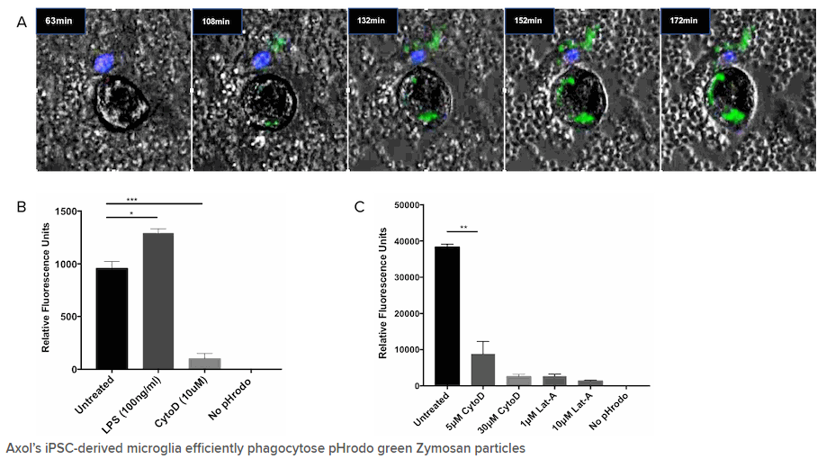
A) pHrodo is a fluorogenic dye that dramatically increases in fluorescence as the pH of the surroundings becomes more acidic, such is the case in the lysosome. pHrodo® Green Zymosan BioParticles® (Life Technologies) were added at 0.5 mg/mL to day 19-microglia plated at 1x10⁵sup>/cm2, and incubated for 3hrs at 37oC, 5% CO2. Nuclei were counterstained with a live Hoechst 33342 stain and live cell imaging was carried out using an on-stage incubator connected to an EVOS Fl Auto imaging system (Life Technologies). Two activated microglial cells can be seen consuming increasing numbers of BioParticles® over time leading greater and more intense fluorescence emission. B) and C) Microglia seeded at 1x10⁵/cm2 (B) or 3x105cm2 (C) were pre-treated with 100 ng/mL lipopolysaccharide (LPS) for 1hr (B), 5, 10μM or 30μM Cytochalasin D (CytoD, phagocytosis inhibitor) or 1μM or 10μM Latrunculin A (Lat-A) for 30mins (B) or 1hr (C) before adding pHrodo® Green Zymosan BioParticles® at 0.5 mg/mL and incubating for 3hrs at 37oC, 5% CO2. Lat-A inhibited phagocytosis more strongly than CytoD, in line with Lat-A being a more potent inhibitor of actin polymerization. Fluorescence was quantified using a SpectraMax iD3 microplate reader (Molecular Devices), and values were blanked against a no-cell control well. N=4 for both experiments, One-way ANOVA *P<0.05, **P<0.01, ***P<0.001

Further Functional Phagocytosis data (courtesy of Medicines Discovery Catapult; Alderley Edge, Cheshire, UK)

Phagocytosis is used as a primary measure of functional Microglia activity. The Cryopreserved Microglia (ax0668) exhibit substrate-dependent phagocytosis when exposed to fluorescent pHrodo labelled beta-amyloid. The Human IPSC derived cryopreserved microglia are shown to be highly phagocytic in nature and equivalent to fresh cells.


Immunocytochemistry of Human iPSC-derived Microglia co-cultured with Human iPSC-derived cortical neurons reveals microglia specific maker expression TMEM119 and neuronal marker β-3 Tubulin.


A co-culture model of neurons, astrocytes and microglial cells were infected with the IncuCyte® NeuroBurst Orange Reagent (a genetically-encoded fluorescent calcium sensor). Calcium-flux kinetics were captured and analysed. Data kindly provided by Sartorius.


관련 자료 

Publications

주문정보

주문정보 - Cat No, PRODUCT, SIZE, 수량 등 항목으로 구성되어있습니다.
  Product Cat.No. Size Maker Qty Data Sheet MSDS
전체보기

추천제품

자료

웨비나/Video

TOP

제품 문의

0.5 ml Elite Pre-stained Protein Ladder (2 x 0.25 ml) PAL-EPL-500 0.5ml 500
Maker
Cat.No.
Product
Size
Qty
Data Sheet
MSDS

로그인

로그인