특징
- Claudin은 tight cell junction을 유지하는 단백질로 다양한 조직에서 발현됩니다. Claudin 18 isoform 2 (CLDN18.2)는 정상시에 위 점막 상피세포에서만 발현되는 단백질이지만, 선암, 췌장암, 식도암, 폐암등과 같은 특정암 발생시 과발현됩니다.
- 제한된 발현으로 인해 CLDN18.2는 위암과 췌장암 치료를 위한 잠재적인 약물 표적이 됩니다.
- Claudin 18.2 (CLDN18.2)를 발현하는 CHO, HEK 세포주 및 단백질과 항체를 제공합니다.
- CLDN18.2와 binding하는 약물 후보의 선별에 이용됩니다.
제품
1. CHO-K1/CLDN18.2 Stable Cell Line (#M00916)
- Clonality : Single clone
- Stability : Stable expression of CLDN18.2 for more than 15 passages
- Culture condition :
- Freeze Medium : 72% F12k, 20% FBS, 8% (V/V) DMSO
- Culture Medium : F12K, 10% FBS, 500μg/ml Geneticin (G418 Sulfate)
2. HEK293/CLDN18.2 Stable Cell Line (#M00917)
- Clonality : Single clone
- Stability : Stable expression of CLDN18.2 for more than 15 passages
- Culture condition :
- Freeze Medium : 49% DMEM, 45% FBS, 6% (V/V) DMSO
- Culture Medium : DMEM, 10% FBS,500μg/ml(G418 Sulfate) Geneticin (G418 Sulfate)
3) MonoRab™ Claudin 18.2 Antibody (126C1), mAb, Rabbit (#A02180)
- Clonality : Monoclonal
- Clone ID : 126C1
- Host Species : Rabbit
- Immunogen : Recombinant Human Claudin 18.2 Protein and DNA
- Conjugate : Unconjugated
- Form : Liquid
- Storage Buffer : Supplied in PBS, pH 7.4, containing 0.02% ProClin300
- Concentration : 1.5 mg/mL
4) CLDN18.2 Protein (#Z03504)
- Species : Human
- Protein Construction : Poly-His CLDN18.2 (Met1-Lys207)
- Accession : P56856-2
- Purity: > 80% as analyzed by SDS-PAGE
- Biological Activity : Immobilized CLDN18.2, His, Human (Cat. No.: Z03504) at 1.0 μg/mL (100μl/well). Dose response curve for anti-Claudin 18.2 Antibody with the EC50 of 13.75ng/mL determined by ELISA.
- Expression System : Insect Cells
- Theoretical Molecular Weight : 27.7 kDa
- Formulation : Supplied as a solution in 50 mM Hepes, 500 mM NaCl, 10% glycerol, 0.2% DDM, 0.04% CHS, pH 7.5.
참고 데이터
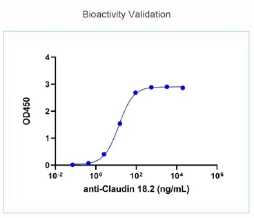
그림 1. Bioactivity of CLDN18.2, hFc, Human (Cat. No.: Z03709) was validated by ELISA. Immobilized anti-Claudin 18.2 antibody was at 1 μg/mL (100 μL/well). Dose response curve for CLDN18.2, hFc, Human with the EC50 of 17.60 ng/ml was determined by ELISA. CLDN18.2, hFc, Human was found to be an active membrane protein.
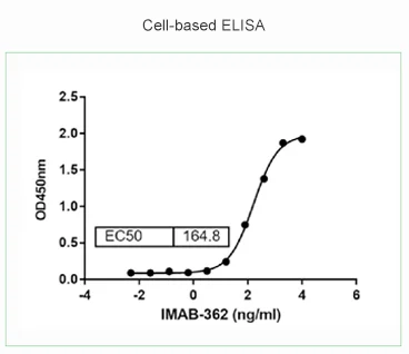
그림 2. Cell based ELISA with CHO-K1/CLDN 18.2 cell line (Cat. No.: M00916). CHO-K1/CLDN18.2 cells were plated at 8x10⁴ cells /well in 100 μl DMEM plus with 10% FBS in 96-well plate overnight. Then the cells were fixed with paraformaldehyde for 15 minutes. After washing, the cells were incubated with gradient concentration of IMAB-362 (anti-CLDN18.2 antibody) for 1 hour, and incubated with goat anti-human IgG (H&L) [HRP] antibody. After incubation with TMB substrate, OD450 was measured by a microplate reader.
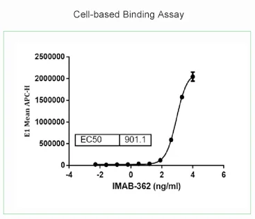
그림 3. Cell-based binding assay with CHO-K1/CLDN 18.2 stable cell line (Cat. No.: M00916). CHO-K1/CLDN18.2 cells were plated at 5x10⁵ cells/well in 100 µL of PBS in 96-well plate, incubated with gradient concentration of IMAB-362 (anti-CLDN18.2 antibody) on ice for 1 hour. The supernatant was discarded and the cells were incubated with goat anti-human IgG antibody at 10 µg/mL on ice for 1 hour. The mean fluorescence intensity of APC (Mean APC-H) was analyzed in live cell gate by a flow cytometer .
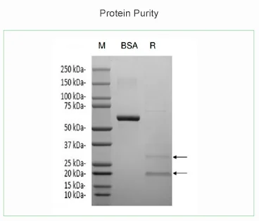
그림 4. The purity of CLDN18.2, His, Human (Cat. No.: Z03504) was assessed by SDS-PAGE. Purity > 90% was observed for 2 μg of protein.
(Lane BSA: 2 μg BSA, Lane R: 2 μg CLDN18.2, reducing (R), no heat treatment before loading.)
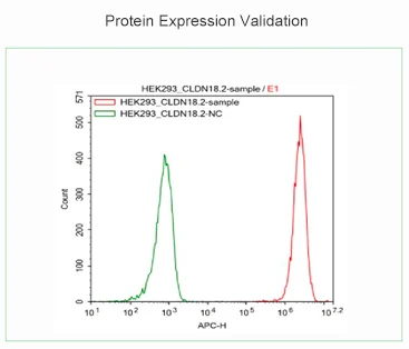
그림 5. Protein expression of CLDN18.2 was validated in HEK293/CLDN18.2 stable cell line (Cat. No.: M00917) using FACS. The FACS analysis of CLDN18.2 expression in HEK293/ CLDN18.2 clone was conducted using anti-CLDN18.2 antibody. The cells analyzed were live cells (E1 gate). (Green line: negative control, treated with goat anti-human IgG antibody. Red line: tested sample, treated with anti-CLDN18.2 antibody.)
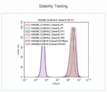
그림 6. Stability of CLDN18.2 expression in HEK293/CLDN18.2 stable cell line (Cat. No.: M00917) was tested using FACS. The stability of expression was tested for cell samples from 20 passages using anti-CLDN18.2 antibody. The cells analyzed were live cells (E1 gate). (Blue line: blank control, untreated cells. Purple line: negative control, treated with goat anti-human IgG antibody. Experimental group: cell samples of different passages.)